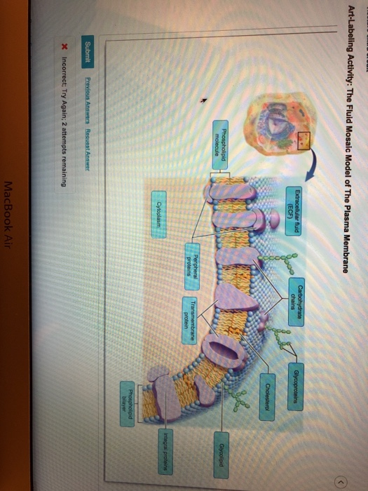
When youve finished answering as many of the questions as you can scroll down to the bottom of the page and check your answers by clicking Score. Glucose a small polar solute uses a membrane transporter a protein carrier to cross the plasma membrane via facilitated diffusion.

Biology Online Journal The Fluid Mosaic Model Of A Cell Membrane Cell Membrane Biology Online Cell Membrane Structure
Choose the best answer from the four options given.
. Diagram Quiz on Fluid Mosaic Model of Plasma Membrane is designed to assess your understanding about the Fluid Mosaic Model of Plasma Membrane. Essential Cell Biology 4th Edition Bruce Alberts Dennis Bray Karen Hopkin. Brain Cranium and Meninges Close-up View of Cranial Meninges Part A Drag the labels to the appropriate location in the figure.
Gas Exchange -- The Respiratory System. Pre-Quiz - Part 2. Use the microscope to identify the nucleus and plasma membrane of cells.
Structures of the Alveoli and the Respiratory Membrane. The Pleurae and Pleural Cavities. Discuss a cells life cycle including the stages of interphase and mitosis.
Functions of antibodies Complement activation Opsonization Mast cell Phagocytic cell Complement protein Precipitation Agglutination Neutraluation Agglutination precipitation FUNCTION K Inflammatory G. In simple diffusion small nonpolar and lipid-soluble substances. Hormones released by the posterior lobe of the pituitary and their target organs.
Instructors may assign this figure as an Art Labeling Activity using Mastering AP Activity 2 Practicing Using Correct Anatomical Terminology Use a human torso model a human skeleton or your own body to practice using the regional and directional terminology. Anterior or posterior 2. Drag the appropriate labels to their respective targets.
The serous membranes of the ventral body cavities Part A Drug the approprate labels to their respective turgels Right ung Left lung Postentur Pustenior Heset Help Coniriue OAsk me anything. Membrane Zona pellucida Sperm Oocyte sperm-binding membrane receptors Granulosa cells of corona radiata 4The sperm forms an acrosomal process which binds to the oocytes sperm-binding receptors. Art-labeling Activities Use the art-labeling activities to quiz yourself on key anatomical structures in this chapter.
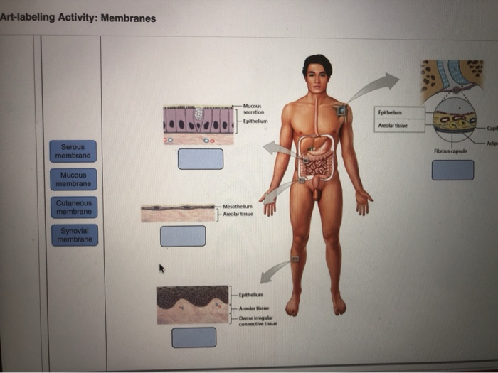
To learn the types of cell junctions. Membranes Label the various types of membranes and the types of secretion associated with each one. The three categories of connective tissues are connective tissue proper cartilage connective tissue and bone connective tissue.
Types of Cell Junctions Learning Goal. 12846 Art-Labcling Acthity The scrous membrancs of she vantral body cavitics Art-Labeling Activity. Part A Drag the label to the appropriate type of membrane or secretion.
Correctly identify the differences between a simple and a compound gland. Structures of the Alveoli and the Respiratory Membrane Match the. Like the dorsal cavity the ventral cavity has two subdivisions.
Membranes 64 of 100. A Structural Classification of Exocrine Glands 19 of 100. Nucleus nuclear envelope nuclear pore plasma membrane cytosol rough Study Resources Main Menu.
Gas Exchange -- Path of Air. View Exercise 4 Review Sheet Art labeling Activity 1 1 of 2png from BIO 168 at Coastal Carolina Community College. 6 Entry of sperm contents tail and plasma membrane remain.
Of the three layers of the meninges the dura mater the arachnoid layer and the pia mater. Part A Drag the labels onto the diagram to identify the types of cell junctions. 5 The sperm and oocyte plasma membranes fuse allowing sperm contents to enter the oocyte.
1159pm on Friday September 22 2017 To understand how points are awarded read the Grading Policy for this assignment. Classifying Epithelia 20 of 100. The popliteal region is _____.
State a function of each organelle. Label the types of cell junctions. Art-labeling Activities Use the art-labeling activities to quiz yourself on key anatomical structures in this chapter.
During which phase of the cell cycle does DNA duplication or replication take place. Meninges are connective tissue membranes that line the neurocranium and vertebral canal and enclose the central nervous system cns brain and spinal cord. Anatomy of the Larynx Drag the appropriate labels to their respective targets.
The connective tissue framework accomplishes all of the listed functions. Art-labeling activities Practice Anatomy Lab PAL virtual anatomy. Brain Cranium and Meninges Close-up View of Cranial Meninges Part A Drag the labels to the appropriate location in the figure.
Location of the major endocrine organs of the body Figure 96. They are flat thin scale-like cells that allow for diffusion of gases.

Honors Biology Lawrenceville Cells Plasma Membrane Cell Membrane Cell Membrane Coloring Worksheet
Solved Art Labeling Activity Membranes Mucous Epithelium Chegg Com

Serous Membrane Large Intestine Health Board

Body Cavity Regions Membrane Large Intestine Body

Art Labeling Activity Membranes Diagram Quizlet
Solved Art Labeling Activity The Fluid Mosaic Model Of The Chegg Com
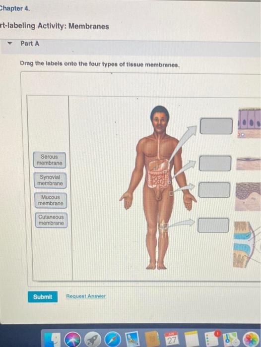
Solved Chapter 4 T Labeling Activity Membranes Part A Drag Chegg Com
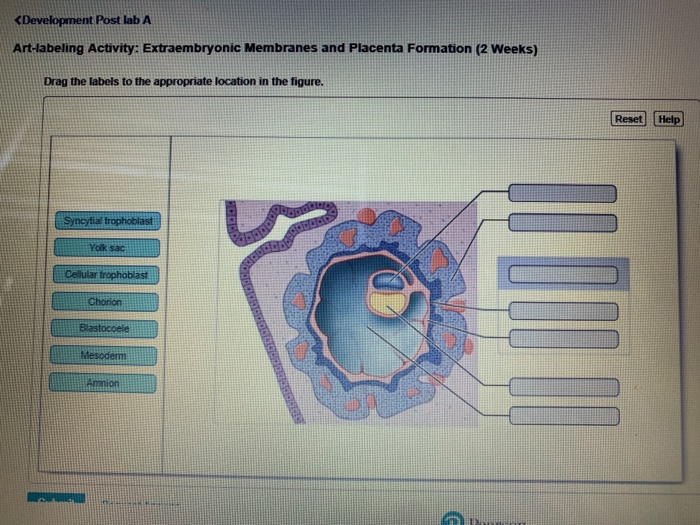
Solved Development Post Lab A Art Labeling Activity Chegg Com
0 komentar
Posting Komentar